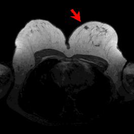
| Fig. 1: MRI mammography of a patient with breast cancer in the upper medial quadrant of the left breast (arrow). |
| Fig. 1: MRI mammography of a patient with breast cancer in the upper medial quadrant of the left breast (arrow). |
|
Focal breast lesions
frequently cannot be classified based on clinical findings and X-ray
mammography alone. In this context, dynamic contrast-enhanced MRI mammography
has been established as a valuable tool for clinical decision making providing
additional insight into regional tissue properties with regard to focal
differences of contrast-agent uptake.
Innovative algorithms for exploratory data analysis can support the clinical interpretation of MRI mammography data by the medical doctor, e.g. by providing useful visualization strategies for multidimensional biomedical image time-series. Figs. 2 and 3 show an example of unsupervised cluster analysis of MRI mammography time-series data obtained from a patient with breast cancer (fig. 1). The procedure is able to segment the lesion from the surrounding breast tissue, as can be seen from clusters #0 and #1 in fig. 2. The corresponding signal time-series in fig. 3 indicate a rapid and strong contrast-agent uptake in the central region of the lesion (cluster #0), surrounded by a semicircular area of slightly reduced signal amplitude (cluster #1). Hence, this visualization strategy can provide a subclassification within the tumour with regard to different contrast-agent uptake patterns. |
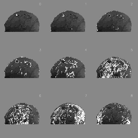 |
| Fig. 2: Cluster assignment maps for MRI |
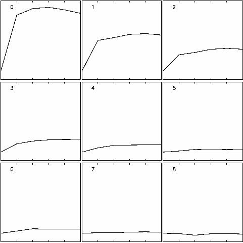 |
| Fig. 3: Corresponding time-series for fig. 2 |
|
Last Update: March-20-2002 |