Segmentation can be defined
as the identification of meaningful image components. It is a fundamental
task in image processing providing the basis for any kind of high-level image
analysis. In medical image processing, a wide range of applications is based on
segmentation, e.g. the volumetric analysis with respect to normal or
pathological organ development, temporal monitoring of size and growth in
pathological processes, or as a basis for the applicability of automatic image
fusion algorithms when combining the complementary information obtained by different
image acquisition modalities.
In our group, we focus on
several segmentation problems that are of specific interest in the field of
neuroradiology, such as high-precision volume measurement of anatomical tissue
classes in normal subjects and patients with psychiatric disorders, or the
temporal monitoring of focal lesions for therapy control in patients with
Multiple Sclerosis.
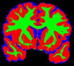
Fig. 1 shows a T1-weighted
MRI cross-section from multispectral MRI data of the brain in a normal
subject. Fig. 2 presents a fully-automatic segmentation of the data with regard
to structure classes Gray Matter (plotted red), White Matter (plotted
green), and Cerebrospinal Fluid (plotted blue). This result has been obtained
by a novel neural network algorithm (so-called Deformable Feature Map)
developed by our group that provides adaptive plasticity in function
approximation problems. It reduces a class of similar function approximation
problems to the explicit supervised one-shot training of a single data set, which
is followed by a subsequent similarity transformation based on a self-organized
deformation of the underlying multidimensional probability
distributions.
|